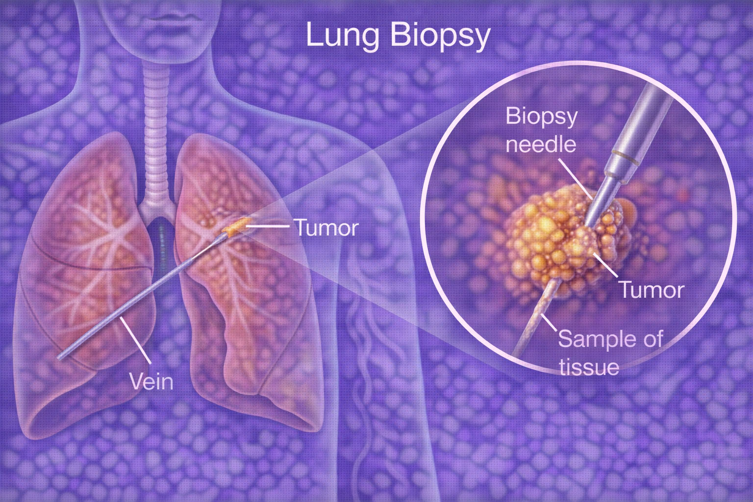
A lung biopsy is a medical procedure in which a doctor takes a small sample of tissue from the lung and sends it to the lab for detailed testing.
A lung biopsy helps to:
- Find out if a lung spot is benign (non-cancerous) or cancerous
- Diagnose infections like TB or fungal disease
- Detect inflammation or scarring diseases
- Choose the right treatment quickly and accurately
Common ways to do a lung biopsy:
- CT-guided needle biopsy (most common)
- Bronchoscopy biopsy (through the airways)
- Surgical biopsy (only in selected cases)
Some lung conditions that are diagnosed through a biopsy also involve abnormal or damaged blood vessels that bleed into the airways, causing a patient to cough up blood. When this happens, bronchial artery embolization is performed to seal the bleeding vessel precisely through a catheter, stopping the bleeding without any chest surgery. In patients with lung cancer, TB, or fungal infections confirmed on biopsy, this is often the most urgent treatment needed before anything else can begin.
A lung biopsy itself can occasionally cause a small amount of fluid to accumulate in the space surrounding the lung — a complication called pleural effusion. This can also develop independently as a result of the underlying lung condition being investigated. When significant fluid builds up and causes breathlessness or discomfort, pleural fluid drainage is performed to remove it safely using image guidance — relieving symptoms and allowing the lung to expand fully so that healing and recovery can progress.
The CT-guided needle technique used in a lung biopsy is part of a broader family of minimally invasive tissue sampling procedures performed across many organs in the body. The image-guided biopsy page covers this full range — including biopsies of the liver, kidney, breast, thyroid, and lymph nodes — explaining what each procedure involves, how patients are prepared, and what to expect during recovery.